


Streptomycesiasis is a fungal lesion that affects only the surface of the nail.To prevent the spread of timely infection, you should know the appearance of nail fungi, because treatment can be achieved faster if treatment begins early in the infection.In most cases, this disease occurs in older people due to weak immunity, but the risk of developing the disease can attract everyone’s attention.There is no single classification of fungal nail damage; in medical practice, habitually distinguishing them in terms of localization and the depth of their penetration.The infection also depends on the type of pathogen.
Types of pathogens and major signs
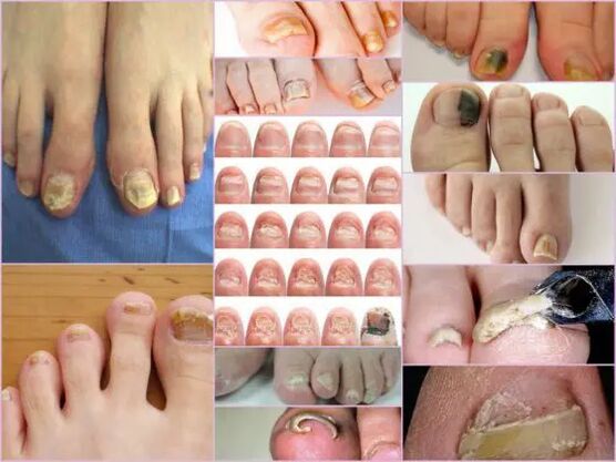
The symptoms of nail fungi in the initial stage are easily determined directly by the nail conditions.This sign is most useful because Onychomycisois always manifests itself in the form of surface color changes, deformation of the bed, exfoliation and any external changes.The latter is expressed through the formation of roughness, grooves and cracks, as well as the spread and generally violating nails.
The main signs of healthy nails are pink and transparency.At any stage, Onychomycosis is characterized by the turbidity of the nail and the change in color to yellow, brown or green black.In the advanced stage, the surface can obtain a black shadow against a background that is almost completely destroyed.
External signs of fungal infection depend on the type of pathogen that is infected.In medical practice, distinguish the following possible lesions:
- Candida is infected with a fungus that is expressed by emitting nails directly at the bottom of the box.Candidiasis in the rollers around the nails will be a characteristic of candidiasis.Complications in this version can have a bacterial source in the form of streptococci or staphylococci, or are expressed in the form of culture medium or psoriasis.
- Skin plants of hairy small capillaries.In this case, the infiltration of infection occurs directly from the free edge of the nail.The first symptom of this pathogen is the appearance of pale yellow dots on the surface.In the tumor field, nails collapse and the spots themselves tend to increase.The common location of tumor positioning is along the plate, parallel to the nail roller, in which case the infection is called the distal lateral.This pathogen is another form of failure - distal, where spots appear in the portions that are removed from the hole, mainly in the middle of the free edge.
Important!
This fungus usually occurs on the thumb of the legs, gradually turning the nails into a loose, pale yellow mass.Without proper treatment, the disease flows into the high nucleus.The nail plates were completely destroyed due to the spread of the surrounding infection.
- Skin plants such as hairy plants.Dinoflavobacteria with this stimulant is also usually based on the frequency of the thumbnails compared to the little fingers of the little finger.The infection of this fungus requires not only the treatment of nails, but also the treatment of the feet, and due to the rapid spread of pathogens.Symptomed diseases can be like Leikonichia, a disease that is common in medical practice.The main signs are white spots that appear in various parts of the nail, and the difference between tumors is non-standard shapes and various sizes.It is easy to distinguish fungi from leaks - in the latter case, the spots are air accumulation and are not observed by fungal damage.
- Mold fungi.This impairment selection is much less frequently compared to candidate or dermatized forms.The main sign of this infection - the surface of the plate is obtained in dark, almost black shadows.Not the entire finger can be completely infected, but part of the nail plate.The first sign of this type of nail fungus on the legs is a drastic change in color.Thyroid disease can be formed in a black or dark green longitudinal form against the background of the rest of the nail.
Diagnosed by change type
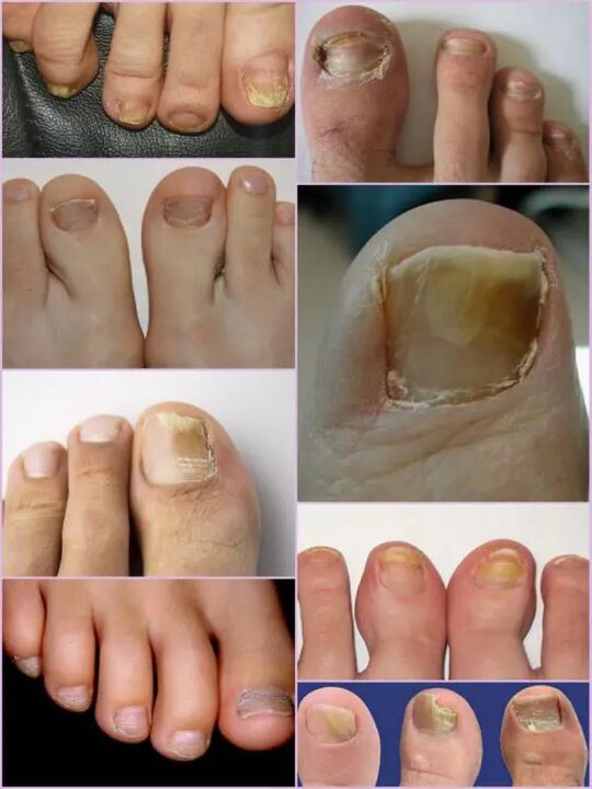
Even in the main stage of the infection, this infection shows positive performance from the first day of the lesion, and it is not difficult to pay attention to nail fungi on the legs.Significant surface deformation and general changes in the condition were observed, rather than the usual light pink nails.The affected area has a matte yellowish hue that appears primarily on the thumb.The type of fungus and the degree of damage are determinants.
In the first stage, fungal damage to the upper leg is the appearance of the small focus.Thickening of the plate and keratinization of the bed beneath the nails are characterized.This stage is accompanied by such phenomena as partial detachment and nail plate emissions, which are the source of infection for healthy people.
Despite the active thickening of the plate, its constant grinding can be observed regardless of the current factors.The characteristics of each stage and the symptoms of the fungus on the legs depend directly on the type of pathogen.
Depending on the changes occurring on the nail plate, there are three options for tumor disease options:
- Thick
- Normal nutrition;
- Shrink.
In the first case, the shadow of the nail plate changes drastically, its damage along the edges and deformation of the surface of the plate.The nails are so thick that they can cause discomfort and pain when walking.The normal nutrition of nails on the legs is distinguished by the presence of a healthy shine, but the plate itself gets some shouting, forming spots on it.There is atrophy type of damage, forming a brown and gray focal point on the nail surface.Thanks to them, it is possible to accurately determine the location of the pathogen.
Important!
The atrophy of nail fungi or a variegated type is characterized by thinning of the nail plate, rather than its growth, and in other cases.Those areas localized to pathogens tend to be isolated.Lack of proper treatment can lead to late stages - total rejection of nail plates.
Localization classification
Another sign that you can separate fungal damage from the toe nails depends directly on where the focus is on the nail plate.This also includes the depth of the pathogen, which in turn allows you to determine the approximate duration of the upcoming treatment and the chances of a quick recovery.
Fungal diseases of the leg nails that locate the location are divided into the following groups:
- Onychomycisois is a white surface type – the appearance of many white spots on the surface of the nail plate.Fungal infections can cause the skin to lose its spotted appearance.Advanced stages can lead to complete destruction and rejection of the board.
- Distal - Develop on the free edge of nails.First, the color change is observed on a small part of the plate, and then the active expansion of its boundaries is followed.The lesions are characterized by pale yellow or brown tones, as well as rough uneven surfaces and gradual exfoliation.
- The developmental stage of lateral tumor disease is similar to the distal end, but is located only on the side of the nail plate.
- All infection - Complete infection of the entire surface;
- Proximal to myelopathy.The disease starts with the inflamed cuticle, and then the fungus quickly affects the nails, and the process itself begins with a small white spot near the inflamed area of the bone roller inflammatory roller.
The most common forms of nails on the legs are transverse and distal, usually caused by skin plants.Impairment forms such as proximal and white can be used as secondary diseases associated with immune system diseases, such as Vich.The total damage to nails with fungi should be considered as an advanced stage of any fungal development under the nail.
Characteristics of fungi under microscope
Despite impressive signs of myomycosis, other diseases associated with skin problems and non-infectivity are accepted due to fungal damage to the leg nails.You can determine the exact diagnosis and type of pathogen in the laboratory only under a microscope, and in the hospital, scratching biomaterials from the diseased area.
The resulting biomaterial was predicted in an alkaline environment and was subsequently added to multiple levels.Studying nail fungi under a microscope allows you to see active pathogens, whose external forms determine their type, distribution scale, and damage level.On the nutrient medium, you can predict approximate growth of colonies, which not only allows for accurate diagnosis but also determines the limitations of infection.
Important!
Since the presence of formicrobial disease can only be determined in the laboratory, you should not postpone the visit to your doctor with slightest suspicion.Nail fungi develop rapidly, and time loss increases treatment.
What is a shocking signal?
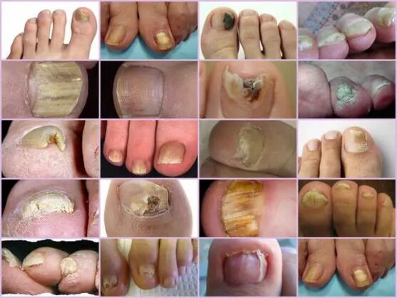
Nail fungi on the legs show certain symptoms that are partially similar to certain skin diseases.The following signs are more likely to indicate thyroxine lesions requiring medical intervention:
- The appearance of yellow spots on the plate, its deformation, structural changes, were not observed earlier.
- The darkening of the plate, the loss of transparency, and some photos of the nail fungus on the legs show a shadow close to black, which is characteristic of the pathogen mold.
- Thickening and keratinizing of the nail plate, and vice versa, is too sharp;
- Reject the fall of nails, scales and debris from the bed;
- The swelling of the roller, hanging it over the plate, releasing liquid;
- Regardless of the type of pathogen, the nails affected by the fungus appear to be inflamed.
All of these symptoms indicate a high risk of infection.Some external symptoms that change the structure of the nail plate may be the result of other diseases.As brittleness increases, the amount of calcium and iron in the body should be increased, and the increase in nodules means any infection in the body, pure white stripes and spots, which may lack copper or zinc.Although in the photo, nail fungus looks like a focal damage to the plate, and the color is closer to yellow, this does not always indicate infection.Shouting can indicate various problems with the stomach and liver.
Although diseases have a wide range of classifications of diseases, it is not difficult to identify thyroidism on the fingers through external signs.However, you can determine the exact type of pathogen, and the stage of damage can only be in the laboratory.